Is computed tomography safe?
One of the most common questions patients ask before undergoing a CT scan is whether the procedure is safe.
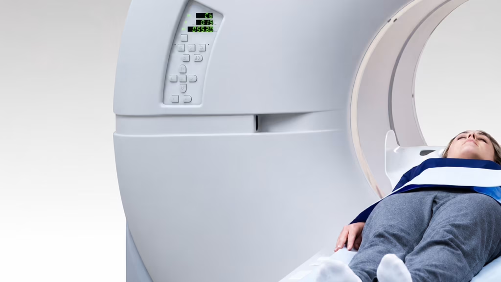
The concern is understandable. Computed tomography uses X-rays to produce detailed cross-sectional images of internal organs and tissues. However, modern CT technology has made this diagnostic method both highly effective and exceptionally safe.
What is the radiation dose during a CT scan?
Modern CT scanners are equipped with automatic dose optimization systems that use only the minimum amount of radiation required to produce high-quality diagnostic images.
The radiation dose depends on the part of the body being examined and the clinical protocol.
Typical doses include:
- Head CT: approximately 1–2 mSv;
- Pelvic CT: approximately 6–11 mSv.
For comparison, everyone is exposed to natural background radiation every day from the environment, as well as additional radiation during long-distance air travel. For this reason, modern CT is considered a safe diagnostic imaging method when clinically indicated.
Contrast-enhanced CT
To provide a more detailed evaluation of blood vessels, internal organs, and tumors, CT examinations are often performed with intravenous contrast enhancement.
This type of examination uses an iodine-based contrast agent that significantly improves diagnostic accuracy.
Before administering contrast, the physician carefully evaluates potential risk factors, including:
- allergic reactions to iodine-based medications or previous contrast agents;
- severe kidney dysfunction;
- certain liver diseases;
- serious cardiovascular conditions.
If any of these conditions are present, the decision to perform the examination is made on an individual basis after carefully weighing the expected benefits against the potential risks.
When may CT be limited?
Although CT is considered a highly safe imaging modality, there are situations in which the examination requires additional evaluation or may be temporarily contraindicated.
These include:
- severe kidney failure, particularly when intravenous contrast is required;
- severe claustrophobia or inability to remain still during the examination;
- certain psychiatric conditions that prevent the procedure from being performed;
- large metallic implants or plaster casts within the area being examined that may affect image quality.